Smart Ephys combines the expertise and experience of three companies: HEKA, Multi Channel Systems, and Warner Instruments. We provide solutions for all areas of electrophysiology from one source. As a trusted partner in your laboratory, we look forward to finding the right solution to support your electrophysiological research.

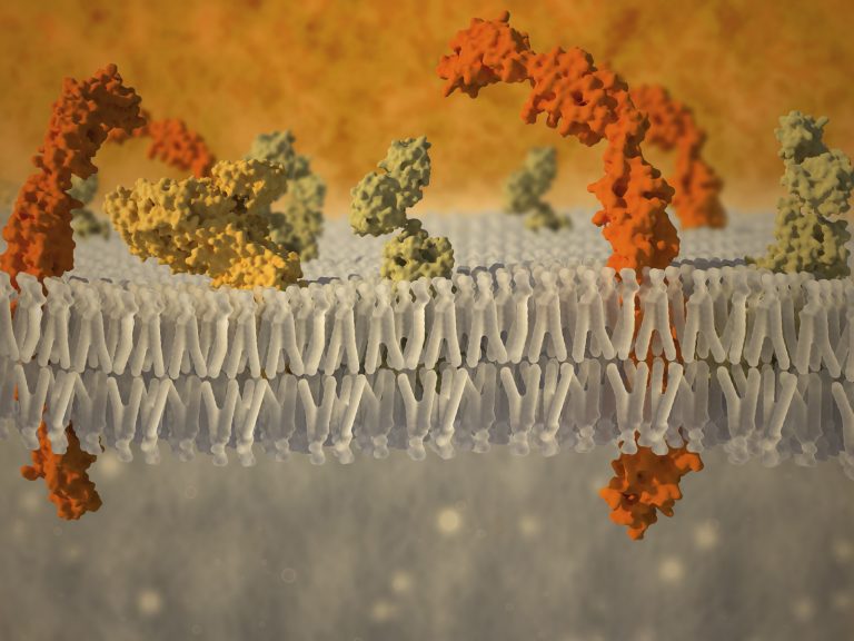
Developed by Nobel Prize winners Erwin Neher and Bert Sakmann, this trusted technique is used in electrophysiological studies of ion channels in tissue sections, individual living cells or patches of cell membrane.

Voltage clamp or current clamp technique is performed in any type of excitable cells, mostly neurons, cardiomyocytes, pancreatic beta cells or muscle fibers. Experiments include slice-recordings, single-cell-layer-recordings, in-vivo-recordings, whole-cell-recordings, and single-channel-recordings.

I am text block. Click edit button to change this text. Lorem ipsum dolor sit amet, consectetur adipiscing elit. Ut elit tellus, luctus nec ullamcorper mattis, pulvinar dapibus leo.

Electrical activity of electrogenic cells and tissues can be recorded by means of microelectrode arrays (MEAs). These arrays allow simultaneous extracellular recording and stimulation from dozens to thousands of recording sites. Since its introduction more than 40 years ago, the technology has been further developed to include different substrates and electrode materials, as well as Multiwell approaches to increase throughput and CMOS based arrays to increase channel count and spatial resolution. We have also innovated a disease specific application of this technology for the study of diabetes using intact Islets of Langerhans.

For a variety of research topics, it is necessary to acquire data from living specimens. Areas including behavior studies, memory functions, or sleep studies require live in vivo data. We offer various platforms for recording and stimulation, tethered or wireless, at low or high channel counts. Depending on your application, experiments may last from minutes to hours or even days; our platforms support all experiment durations.

Ussing chamber systems are critical tools for studying epithelial transport in diverse research areas including genetic disorders, drug discovery and testing, animal nutrition and physiology, the bacterial toxicology.

Scanning Probe Microscopy

Scanning Probe Microscopy (SPM) is a branch of microscopy that forms micrograph images of surfaces or interfaces using a physical probe that scans specimen locally and senses molecules specifically. Electrochemical SPM imaging techniques, not only possesses the advantages of working in non-contact and non-destructive modes, but also may be combined  with a wide arrays of cutting edge microscopy and spectroscopy techniques, such as electrochemical, mechanical, optical, photoelectrical, and fluorescence optical measurements.  The areas of applications range from physical, material to biological science.